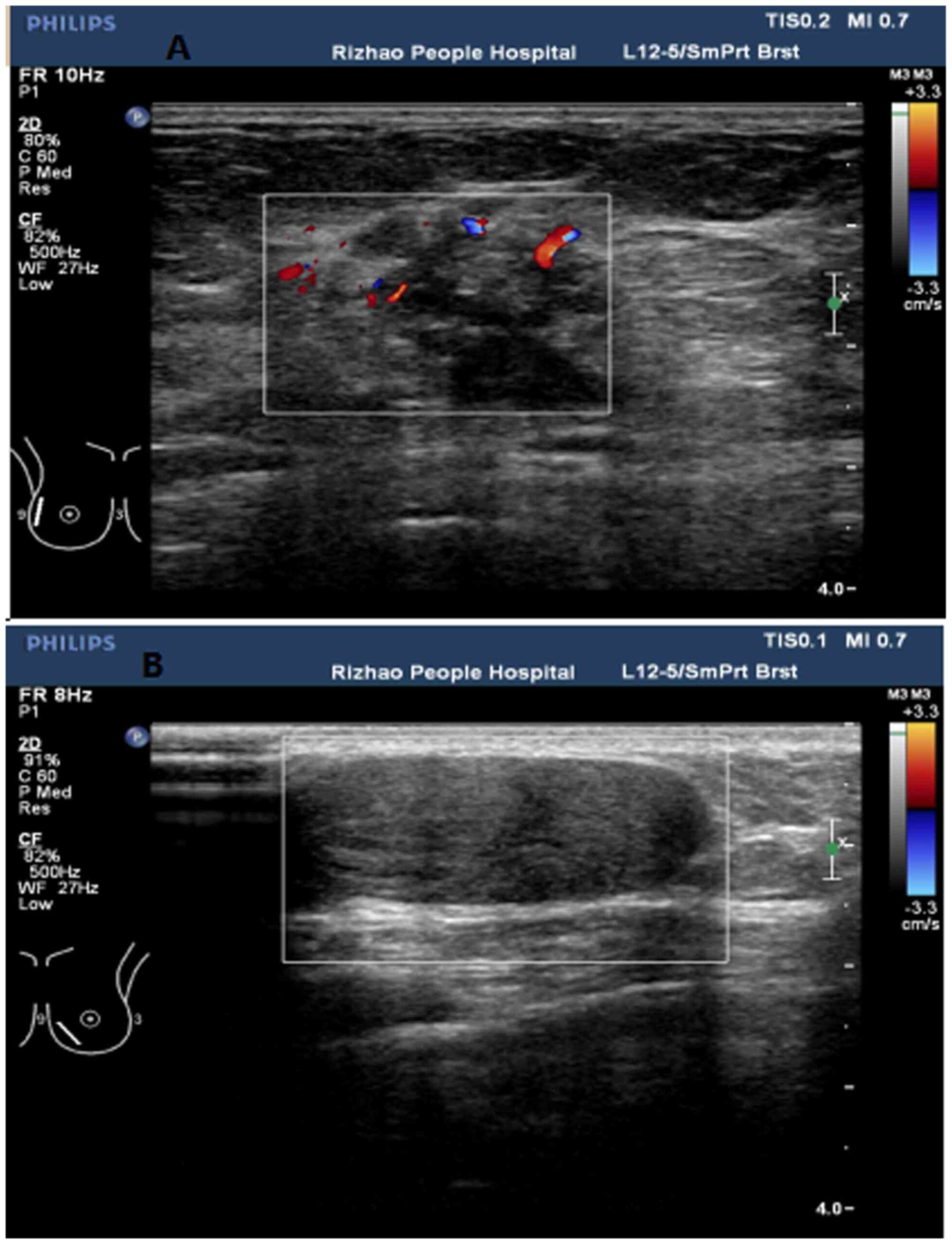
Ultrasound imaging has revolutionized the field of diagnostics, providing a non-invasive window into the physiology and pathology of the human body. One of the most compelling aspects of ultrasound technology is its ability to portray information through a vibrant and intricate palette of colors. These colors, particularly as seen on Doppler ultrasound scans, are not mere decorations; they are the language of our internal systems, conveying a wealth of information about blood flow, tissue characteristics, and even potential abnormalities. Understanding what these colors signify can illuminate the intricate dance of life occurring within us.
At the heart of Doppler ultrasound lies the principle of sound waves interacting with moving structures, specifically blood cells. As the ultrasound probe emits these sound waves, they bounce off the red blood cells and return to the transducer. The varying frequency of these returning sound waves, as they encounter moving blood, codes vital information through color designations, often seen in a mapped format on the screen.
1. The Palette of Color: A Spectrum of Meaning
In the vibrant spectrum conjured by a Doppler ultrasound, each hue tells a compelling story. Color maps can vary between machines, but the underlying principles remain constant. Typically, red indicates structures moving toward the transducer, while blue signifies those moving away. These intuitive color codings allow for immediate visual interpretation, enhancing the diagnostic process.
Imagine the contrast; red reflects vibrancy, pulsating with energy, while blue provides a soothing backdrop, indicative of a retreating force. This dichotomy resets the narrative of blood movement within the body—a dynamic ballet of inflow and outflow, ever in balance.
2. The Significance of Motion: Assessing Blood Flow
But what does it mean when those colors begin to dance erratically or shift unexpectedly? Flow patterns are critical in assessing vascular health. The velocity of blood flow can lead to insights that very well could alter treatment modalities. A bright red hue might suggest high-velocity flow, often seen in arteries supplying oxygen-rich blood to vital organs. Conversely, a muted blue might indicate sluggish flow, a potential harbinger of vascular complications.
In diagnosing issues like deep vein thrombosis (DVT) or carotid artery stenosis, the ultrasound colors reveal telling signs of alterations in blood dynamics. An evolved understanding of these color indicators can transform a perplexing case into a straightforward diagnosis, guiding clinical decisions with precision.
3. The Importance of Context: Beyond Colors
To truly grasp the significance of color in ultrasound imaging, it is crucial to marry these visual cues to their anatomical contexts. For instance, the same hue could elicit different implications when observed in various locations. While red in the heart’s valves might represent healthy flow, in a peripheral artery, it could signify an arterial blockage. Thus, the color spectrum cannot be interpreted in isolation but must be contextualized within the realm of anatomical and physiological knowledge.
4. Understanding Abnormalities: What Colors May Reveal
As we delve deeper, the colors on an ultrasound can become a differential diagnosis tool, opening doors to previously hidden pathologies. The appearance of unexpected colors can indicate various disorders. For example, the presence of a spectral broadening—a widening of the color band—might suggest turbulence caused by a vascular obstruction or a significant valve disorder. Whether marbled with dark patches or illuminated with vibrant brights, each nuance heightens the vigilance of the diagnostician.
Moreover, subtle shifts within these colors can herald the onset of conditions such as arteriosclerosis or turbulent blood flow from aneurysms. Recognizing these variations can mean the difference between life-saving interventions and unfortunate outcomes.
5. Future Directions: Advancements in Ultrasound Technology
Innovation is ever afoot in the realm of ultrasound. With advancements in technology and imaging techniques, the interpretation of color in ultrasound is poised to become even more sophisticated. Machine learning and artificial intelligence are beginning to play pivotal roles in refining color Doppler interpretation, mapping blood flow with unprecedented accuracy. The canvas of ultrasound will thus expand further, tracing the patterns and anomalies of the human body with greater finesse.
As imaging techniques evolve, so too does our understanding of the physiological narratives these colors represent. The future may witness a renaissance in diagnostic capabilities—turning vivid colors into even more compelling stories of health, disease, and the human journey.
6. Conclusion: The Metaphorical Tapestry of Life
With every ultrasound colored display, we are treated to a metaphorical tapestry of life, woven with threads of red, blue, green, and yellow. These hues transform into narratives that articulate the rhythms of our existence; they echo tales of wellness or forewarn of disturbances. The art of interpreting these colors lies not just in recognizing the shades but in understanding their implications for human health.
In essence, Doppler ultrasound does more than merely produce images; it unfurls a visualization of the body’s inner workings, capturing the flow of life in a vibrant tableau. As we sharpen our understanding of what these colors convey, we become not just observers but active participants in deciphering the sophisticated language of our own physiology.