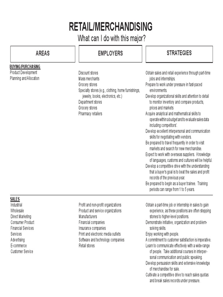
Ultrasound technology, akin to a maestro conducting an orchestra of sound waves, possesses a remarkable ability to illuminate the intricacies of the human body. Among the myriad of colors displayed in a Doppler ultrasound scan, red and blue emerge as the most prominent hues, transcending mere aesthetic appeal to convey vital physiological information. Understanding the significance of these colors is akin to decoding a beautiful yet complex symphony. Herein, we delve into the meanings behind the red and blue on a Doppler ultrasound, unraveling the tapestry of blood flow and its dynamic performance within the vascular system.
At its core, Doppler ultrasound is a non-invasive diagnostic tool that harnesses the principles of echolocation. By emitting high-frequency sound waves that bounce off moving blood cells, this technology allows medical professionals to visualize and measure blood flow dynamics. The resulting color-coded images are not arbitrary; they are a sophisticated language wherein each color speaks volumes about the direction and speed of blood movement.
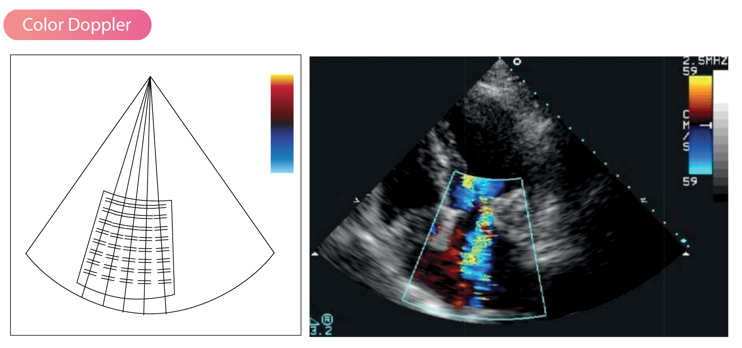
The color red, often associated with warmth and vitality, represents blood flowing toward the ultrasound transducer. Picture this blood as a bright cherry, rushing with vigor to supply organs with the much-needed oxygen and nutrients. The transducer, positioned strategically on the skin, captures these sanguine currents, revealing both the velocity and direction of flow. In the realm of assessed arteries, a rich crimson hue signifies optimal perfusion, heralding a healthy circulatory system and the graceful passage of life through the body’s vessels.
Conversely, blue is evocative of serenity, yet in the parlance of Doppler ultrasound, it conveys a different story. It denotes blood coursing away from the transducer. Imagine this blood as a tranquil azure river, flowing outward to return vital carbon dioxide and cellular byproducts back toward the lungs and heart for rejuvenation. In many cases, blue indicates venous return, an essential journey that completes the cycle of circulation. The aesthetic contrast between red and blue serves not only to captivate the eye but also to elucidate the dance of life within our vascular pathways.
The allure of these colors is further magnified by the Doppler Effect—a phenomenon that has captivated scientists for centuries. Named after the Austrian physicist Christian Doppler, this principle explicates how sound waves shift in frequency relative to the observer’s position. When blood cells approach the transducer, the frequency of the sound waves increases, manifesting as red on the ultrasound imagery. Conversely, as blood cells recede, the frequency diminishes, producing a blue hue. This phenomenon offers a dynamic portrayal of blood flow, allowing practitioners to assess not just direction, but also velocity—critical parameters for diagnosing various medical conditions.
When assessing the implications of red and blue on a Doppler ultrasound, it is crucial to consider the context in which these colors manifest. Various conditions could alter this seemingly straightforward color scheme. For instance, certain heart anomalies might disrupt normal blood flow, resulting in atypical color patterns. A mixture of red and blue in the same region may signal turbulence, indicative of potential vascular malformation or pathology. Such complexities illustrate that while red and blue provide immediate visual cues, the entirety of the ultrasound image must be scrutinized to grasp the full clinical narrative.
Beyond the technicalities lies a profound clinical significance. Doppler ultrasound is instrumental in the diagnosis and management of several medical conditions. Physicians often rely on this technology to detect arterial blockages, assess venous insufficiency, and evaluate cardiovascular health. The colors red and blue essentially become the brushstrokes of a portrait depicting vascular health, guiding clinical decisions that can profoundly affect patient outcomes.
Moreover, the utility of red and blue extends beyond mere diagnostics. They are entrenched in the realm of non-invasive monitoring—an essential component in scenarios such as pregnancy. Obstetric ultrasonography employs Doppler techniques to monitor fetal blood flow, providing insight into the well-being of both mother and child. Understanding the flow dynamics represented by red and blue ensures that healthcare providers can intervene proactively when necessary, establishing a safety net for the developing fetus.
However, in this world of colors, interpretations may vary. The algorithms used to assign red and blue hues may differ among machines, potentially leading to discrepancies in readings across different devices. This variability underscores the importance of skilled interpretation and thorough knowledge of the underlying physiology, positioning experienced clinicians as pivotal players in deciphering and integrating these colorful displays into holistic patient care.
As we navigate through the richness of Doppler ultrasound, it is apparent that the colors red and blue are not mere embellishments but rather the beats and measures of the cardiovascular symphony. They weave together a narrative of health that is as informative as it is vital. While modern medicine continues to advance, with technologies evolving and improving in precision, the fundamental relationship between sound, color, and blood flow remains a captivating aspect of medical diagnostics. Thus, every time a transducer glides over skin, the vibrant dance of red and blue unfolds once more, a reminder of the life coursing beneath, where every pulse tells a story worth listening to.