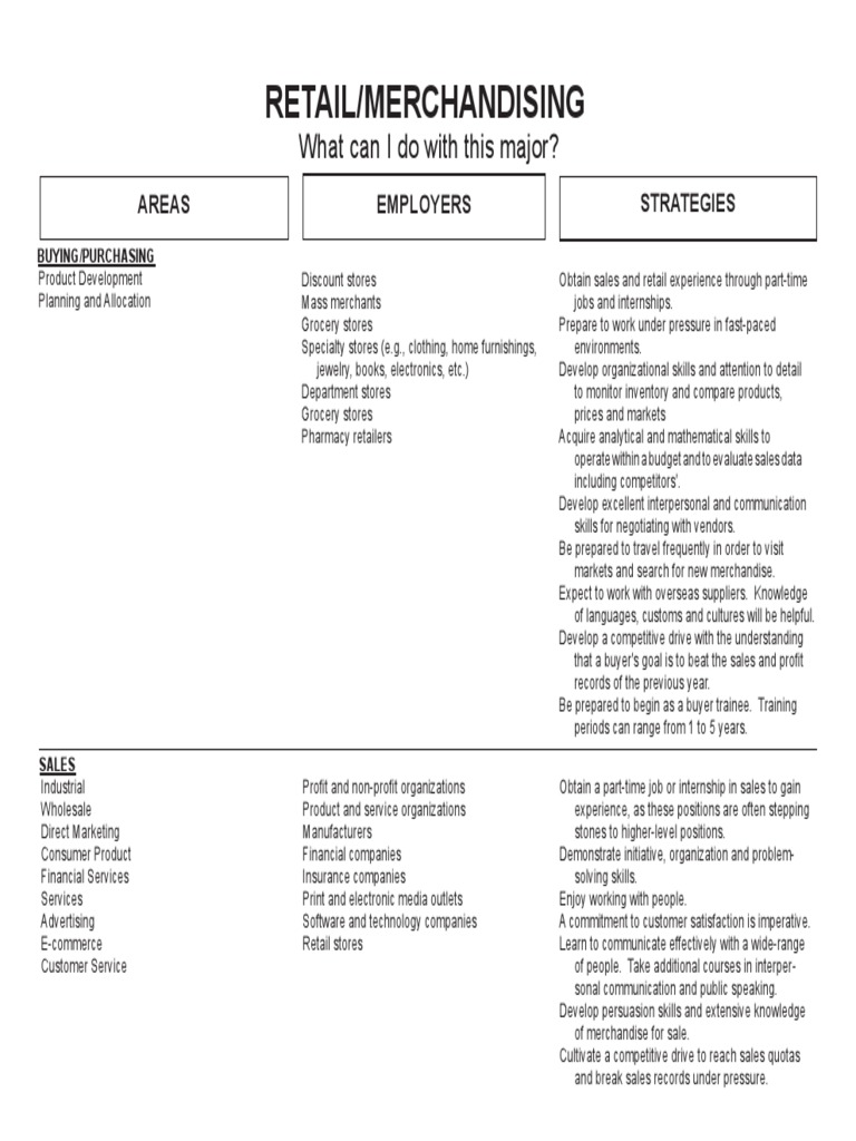
Ultrasound imaging has revolutionized the fields of medicine and diagnostics, providing clear visualizations that allow healthcare professionals to assess various conditions with ease. Among the many techniques utilized in ultrasound imaging, Doppler ultrasound stands out for its ability to measure and illustrate blood flow and the movement of tissues. One of the most captivating aspects of Doppler ultrasound is the use of color to represent different velocities and directions of blood flow. This article delves into the significance of the colors produced during a Doppler ultrasound scan, ensuring a comprehensive understanding of what these colors convey.
The Basics of Doppler Ultrasound
Before we embark on a detailed exploration of color representations in Doppler imaging, it is essential to understand the fundamental principles behind Doppler ultrasound. This technique employs high-frequency sound waves, emitted and received by a transducer, to produce images of the inside of the body. As these sound waves encounter moving objects—such as red blood cells—they reflect off them, causing a change in frequency known as the Doppler effect. This frequency shift is then translated into visual data that can be color-coded for better interpretation.
Interpreting the Color Palette
The colors used in Doppler ultrasound are not merely aesthetic; they serve as a critical tool for diagnosis. Typically, a color Doppler ultrasound utilizes a spectrum of hues to present information regarding the speed and direction of blood flow. Commonly seen colors include red, blue, and various shades in between. Each color indicates a specific flow behavior, allowing for quicker and more intuitive diagnoses.
Red: An Indication of Forward Flow
Red often denotes blood flowing toward the transducer—essentially indicating forward flow. This is particularly important in assessing cardiac function and the efficiency of blood circulation. A bright red hue indicates a high-velocity flow, which may suggest normal or vigorous activity, while a softer red may represent a slower velocity. Understanding these variations can help diagnosticians detect abnormalities such as stenosis or other vascular issues.
Blue: Blood Flow Away from the Transducer
Conversely, blue is indicative of blood moving away from the transducer. This color coding is crucial for evaluating various conditions in both arteries and veins. Like red, varying shades of blue can signify different velocities; a deeper blue may indicate a substantial flow away from the probe, suggesting potential concerns or the need for further investigation. This contrasting coloration between red and blue provides a straightforward visual cue, facilitating immediate recognition of blood flow dynamics.
The Green Spectrum: Transitional Velocities
While red and blue are the primary colors employed in Doppler imaging, shades in the green spectrum often represent transitional velocities. Specifically, these colors may indicate a lower flow rate, or transitional flows that could be characteristic of specific types of blood vessels, such as veins. Physicians utilize this information to gauge the baseline functionality of these vessels and identify anomalies like turbulence, which may be indicative of pathological states.
Understanding Color Aliasing
One phenomenon that one might encounter during Doppler imaging is known as color aliasing. This occurs when blood flow velocities exceed the scale set within the imaging parameters, resulting in an unexpected reversal of color—what should be represented as red may suddenly appear blue and vice versa. Recognizing this occurrence is vital for accurate interpretation, as it can lead to misdiagnosis if overlooked. Technicians must adjust settings to accommodate higher velocities, ensuring that the full spectrum of flow dynamics is captured.
Clinical Applications of Doppler Ultrasound
The implications of Doppler ultrasound span a wide range of clinical applications, making it a relevant tool across various medical specialties. Cardiologists rely on this imaging technique to evaluate heart function, monitor valvular integrity, and assess congenital heart defects. Additionally, obstetricians employ color Doppler imaging to examine placental blood flow and fetal wellbeing, providing vital information that could alter management strategies during pregnancy.
A Broader Fascination with Color in Medical Imaging
Beyond the clinical implications, the use of color in Doppler ultrasound raises intriguing questions about human perception and the symbolic nature of colors. The vibrant red and blue hues can evoke an emotional response, stirring feelings that range from apprehension to fascination. This fascination underscores the broader human interest in visual representation, particularly when the stakes involve health and wellbeing. Color acts as a bridge between the biological and emotional realms, imbuing scientific data with a layer of accessibility that resonates with both practitioners and patients alike.
Final Thoughts: The Convergence of Art and Science
Ultimately, Doppler ultrasound serves as a quintessential example of the convergence between art and science. The colors utilized within these images are not just mere reflections of data; they encapsulate intricate physiological processes and enhance our understanding of the human body. As technology continues to advance and imaging techniques become increasingly sophisticated, the role of color in diagnostics will likely evolve, providing ever-deeper insights into health and disease. Appreciating the nuanced meanings behind these colors can lead to a greater respect for the marvels of medical imaging and its powerful impact on patient care.